
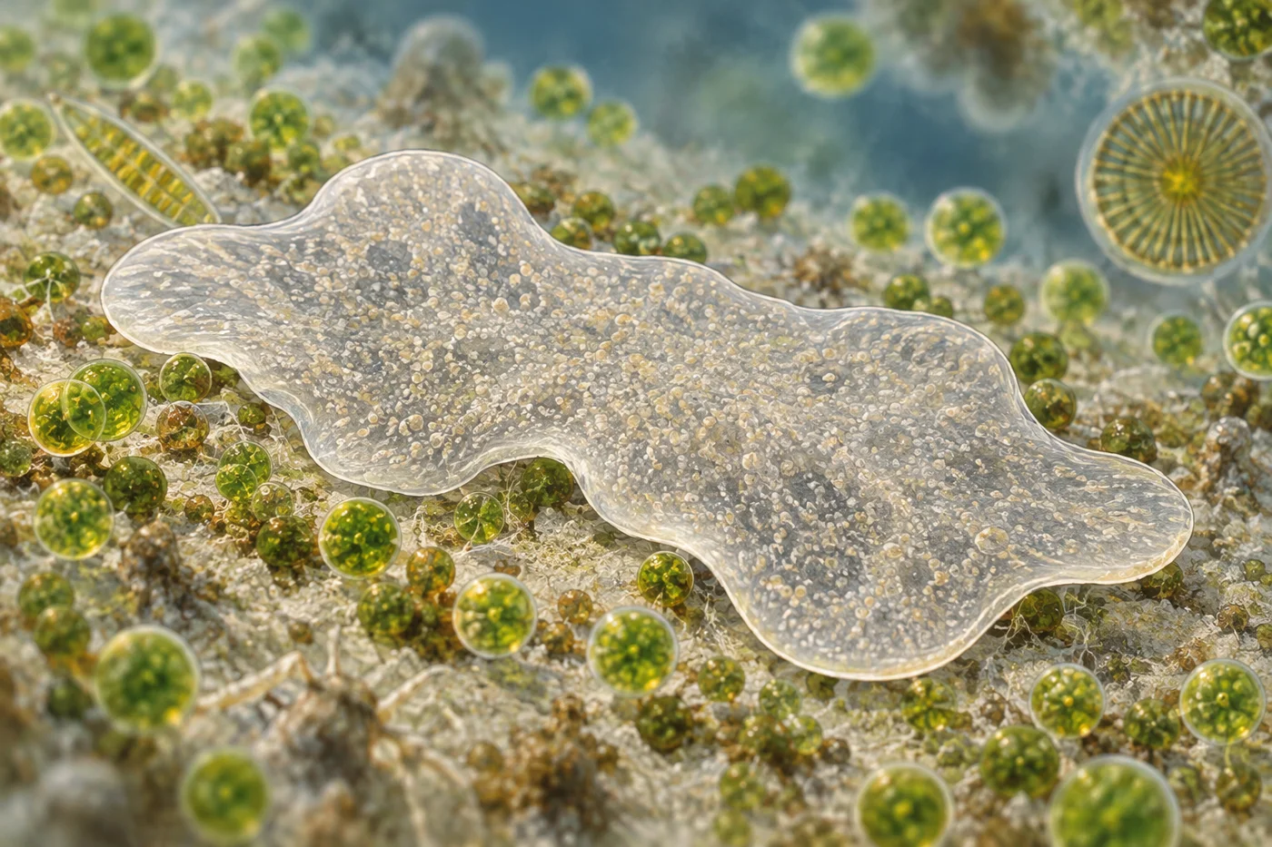
A nervous system is one way to coordinate an animal, but it is not the only way. Our article on animals that move without a brain explores that broader puzzle. Trichoplax adhaerens brings it right down to the dinner table: this tiny marine animal has no mouth or internal digestive cavity, yet it can stop over food, break it down and absorb the results.
Its appearance hardly prepares you for that sequence. A living Trichoplax resembles an irregular, flattened, translucent patch. The outline changes as it moves. Instead of arranging feeding around a tube with an entrance and exit, it uses the narrow space between its lower surface and the surface underneath it.
A flat body with a distinct upper and lower side
Trichoplax belongs to the placozoans, a group of small marine animals with unusually simple-looking bodies. “Simple-looking” is the useful qualification. A disc without a recognizable head can still contain different cells performing different jobs.
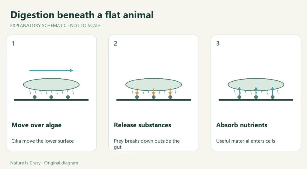
The lower surface carries cilia, tiny moving cellular projections that help propel the animal across a substrate. Food such as microscopic algae lies on or near that substrate. As the animal passes across it, feeding can happen underneath the body. The meal is therefore outside an internal gut but within a small space that the animal’s position creates.
Research on a related placozoan, Hoilungia hongkongensis, revealed more cell diversity than the plain body outline suggests. That finding should not be used to assign every cell type in Hoilungia to Trichoplax. It does show why appearance alone is a poor measure of biological complexity.

Digestion happens beneath the animal
The basic feeding arrangement is external digestion. Rather than swallowing an intact algal cell into a stomach, the animal releases substances into the space beneath it, where prey can be disrupted. Nutrients then become available to cells on the feeding surface.
A live-imaging study of coordinated feeding followed this process as it happened. Animals paused over food, and specialized secretory cells close to prey discharged their contents. The observations connected a local cellular event with a response involving the animal’s wider movement.
The pause is part of the explanation. If a moving body immediately passed beyond the material it had begun to digest, the benefits of secretion could be lost. Remaining over a patch of food makes the arrangement more effective. That is a physical interpretation of the observed behavior, rather than evidence that the animal consciously decides to wait.
“No mouth” also does not mean “no boundary.” The lower body surface still separates cells from their surroundings. Digestion alters food outside that boundary; uptake moves useful material across it. Keeping those steps separate prevents the process from sounding like an animal simply dissolving everything it touches.
Secretory cells supply the machinery
More recent work has examined the cells responsible for secretion in greater detail. A study combining molecular markers, microscopy and observations of living animals identified secretory cell types implicated in feeding, defense and behavioral regulation. The researchers found different cell types contributing to prey breakdown, rather than one general-purpose cell doing every part of the job.
This approach matters because a cell can be described in several ways. Its shape may be visible under a microscope; its expressed genes may suggest the substances it makes; live observation may show when it releases those substances. Agreement between these kinds of evidence gives a stronger functional explanation than any one alone.
External processing has parallels elsewhere in nature, although the structures differ enormously. For example, hoatzin digestion relies on microbial processing within an enlarged part of the digestive tract. Comparing the two helps distinguish a shared problem—making food usable—from the very different locations and partners involved.
Coordination does not require ordinary synapses
Trichoplax lacks the familiar arrangement of neurons connected by synapses. Nevertheless, its cells can respond to signals and its body can change behavior. Absence of a conventional nervous system is not absence of communication.
The secretory-cell research identified cells expressing short peptides and other products that may contribute to signaling. The precise roles of all these cell types are still being investigated. It would go beyond the evidence to imagine a fully mapped substitute brain hidden inside the animal.
A useful way to read the feeding observations is to keep the scales separate. A nearby secretory cell acts locally; other cells participate in movement or absorption; the whole animal remains over the food patch. The research question is how those responses become coordinated. Naming the overall result does not by itself reveal every signal linking the steps.

Other cells clean up and repair damage
The body also contains fiber cells with branching processes. A study of their functions found evidence that these cells engulf bacteria and cellular debris and participate in wound healing. The authors described them as macrophage-like, referring to similarities in function rather than claiming that a placozoan has the same immune system as a person.
Those results add an important dimension to the feeding story. An animal working directly against a surface must also maintain its own tissues. Processing food, responding to unwanted material and repairing injury are different tasks, even when the body carrying them out has no familiar organs.
Why this tiny animal changes the question
Trichoplax does not demonstrate that organs or neurons are unnecessary for every kind of animal life. Its feeding method works within a particular body size, shape and environment. What it demonstrates is that an animal can organize a successful feeding sequence without the mouth-to-stomach route we usually imagine.
For another unconventional arrangement of animal tissue and supporting structures, see how glass sponges build their skeletons. The lesson from Trichoplax is more specific than “tiny animals are strange”: familiar-looking organs are only one possible way to divide the work of living.
Featured image credit: AI-generated editorial illustration of Trichoplax. Conceptual imagery, not a documentary photograph; anatomy and scale are illustrative.