In 1992, researchers investigating a pneumonia outbreak in Bradford, England, collected a water sample from a cooling tower. Under a light microscope, they saw a peculiar microbe, a speck large enough to be seen alongside bacteria. Following standard lab procedures, they applied a Gram stain, and the particle turned purple, indicating it was Gram-positive, a classic bacterial trait. For over a decade, this mysterious entity was labeled “Bradfordcoccus,” filed away as a stubborn, unculturable bacterium, and largely forgotten. This simple misidentification held the secret to a new class of biological entities, the giant viruses, which would soon force scientists to reconsider the very definition of life.
The Accidental Discovery of Giant Viruses
The story of Bradfordcoccus could have ended there, a footnote in a dusty lab notebook. But in 2003, a team of French researchers led by Jean-Michel Claverie and Didier Raoult decided to take another look. Their curiosity was piqued by the microbe’s refusal to be categorized. Unable to sequence a key ribosomal gene that is universally present in bacteria, they suspected something was amiss. They turned to a more powerful tool: the electron microscope.
What they saw was not a bacterium at all. Instead of a simple cell, the microscope revealed a particle with a distinct 20-sided icosahedral shell, the unmistakable signature of a virus. Yet, its scale was staggering. It was a behemoth, dwarfing every known virus and even rivaling some small bacteria in size. This was no ordinary pathogen. It was something entirely new.
The researchers renamed it Mimivirus, short for “mimicking microbe,” a nod to how effectively it had masqueraded as a bacterium for so long. The mimivirus discovery was more than just finding a big virus. It was a scientific bombshell that sent shockwaves through the fields of virology and microbiology. For a century, viruses had been defined by what they were not: they were not cellular, they were not visible under a light microscope, and they were not genetically complex. Mimivirus violated all these rules. It was so large and intricate that it blurred the line between the simple, inert particles viruses were thought to be and the complex, living world of cells. The discovery opened a floodgate, revealing that an entire class of viral titans had been hiding in plain sight, simply because no one thought to look for something so paradoxical.
Shattering the Boundaries of the Viral World
Before Mimivirus, the definition of a virus was clear and concise. They were known as “filterable agents,” a term born from 19th-century experiments where infectious fluid was passed through porcelain filters fine enough to trap all known bacteria. The invisible agent that passed through was a virus. This defined them by their minuscule size and their fundamental simplicity. Viruses were considered biological artifacts, existing at the edge of life. They lacked cellular structures, had no metabolism, and most importantly, they did not have ribosomes, the cellular factories required to build proteins. They were obligate parasites, needing to hijack a host cell’s machinery to do anything at all.
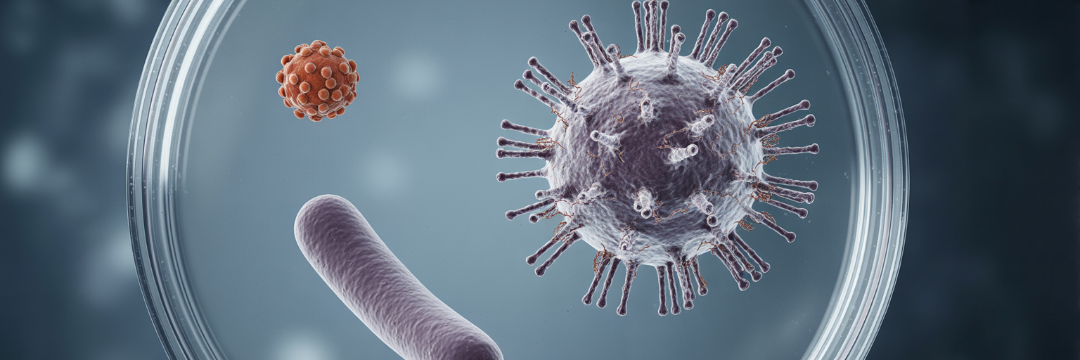
A typical poliovirus measures about 30 nanometers across, while an influenza virus is around 100 nanometers. In contrast, the Mimivirus capsid, or protein shell, is about 500 nanometers in diameter. With a dense layer of protein filaments extending from its surface, its total size reaches nearly 750 nanometers. To put that into perspective, if a common cold virus were the size of a tennis ball, a Mimivirus would be as large as a small car. This makes it easily visible with a standard light microscope, the very reason it was first mistaken for a bacterium. It occupies a biological gray zone, smaller than most bacteria like E. coli (around 2,000 nanometers long) but vastly larger than any virus previously characterized.
This section helps answer the question of what are giant viruses by illustrating their scale. Upon infecting its host, a single-celled amoeba, a giant virus doesn’t just inject its DNA. It constructs a large, distinct compartment inside the host cell known as a “virus factory.” This structure becomes the central hub for producing new virus particles, coordinating the assembly of viral components in a way that seems almost autonomous. This complex behavior further challenges the old view of viruses as passive parasites.
| Feature | Conventional Virus (e.g., Influenza) | Giant Virus (e.g., Mimivirus) | Bacterium (e.g., E. coli) |
|---|---|---|---|
| Average Size | ~100 nanometers | ~750 nanometers | ~2,000 nanometers |
| Visibility | Electron Microscope Only | Light Microscope | Light Microscope |
| Genome Size (Base Pairs) | ~13,500 | ~1,200,000 | ~4,600,000 |
| Ribosomes | Absent | Absent | Present |
| Independent Metabolism | Absent | Absent | Present |
A Genome That Rivals a Bacterium’s
If the size of Mimivirus was shocking, its genetic blueprint was revolutionary. Viruses were long considered models of genetic efficiency, carrying only the bare-minimum instructions needed to replicate. The HIV genome, for example, contains just nine genes, while the influenza virus has about fourteen. They are masters of doing more with less.
The Mimivirus genome, however, is anything but minimal. It contains approximately 1.2 million base pairs of DNA, encoding for over 900 genes. This is larger than the genomes of dozens of free-living bacteria and archaea. It was as if scientists had found a bicycle with the instruction manual for a spaceship. The contents of this manual were even more surprising than its size.
Among its hundreds of genes, researchers found instructions for functions no one ever expected to see in a virus. These included genes for:
- DNA repair enzymes to fix its own genetic code.
- Sugar, lipid, and amino acid metabolism.
- Aminoacyl-tRNA synthetases, enzymes that play a critical role in translating genetic code into proteins.
The presence of translation-related genes was particularly stunning. This machinery was considered a strict hallmark of cellular life, one of the key dividing lines between viruses and living organisms. Finding these genes in a virus was like finding a fish with rudimentary lungs. A study in PLOS Genetics on ‘Translation in Giant Viruses’ highlights their unique mix of bacterial and eukaryotic features, underscoring the unexpected genetic capabilities these entities possess. Furthermore, a significant portion of the Mimivirus genome consists of “orphan genes,” or ORFans, which have no known homologs in any other life form on Earth. This deepens the mystery of their evolutionary origins. This genetic complexity suggests a history far more intricate than that of a simple parasite. The study of these disturbing parasite life cycles you wont forget reveals how evolution can produce highly sophisticated genetic toolkits for survival, and giant viruses appear to be another prime example.
The Expanding Family of Viral Titans
The discovery of Mimivirus was not an isolated event. It was the first clue that an entire undiscovered branch of the viral world existed. Scientists, now knowing what to look for, began a “gold rush” to find other giant viruses, focusing their search on the single-celled amoebae that serve as their primary hosts. What they found was a stunning diversity of viral titans, each with its own unique characteristics.
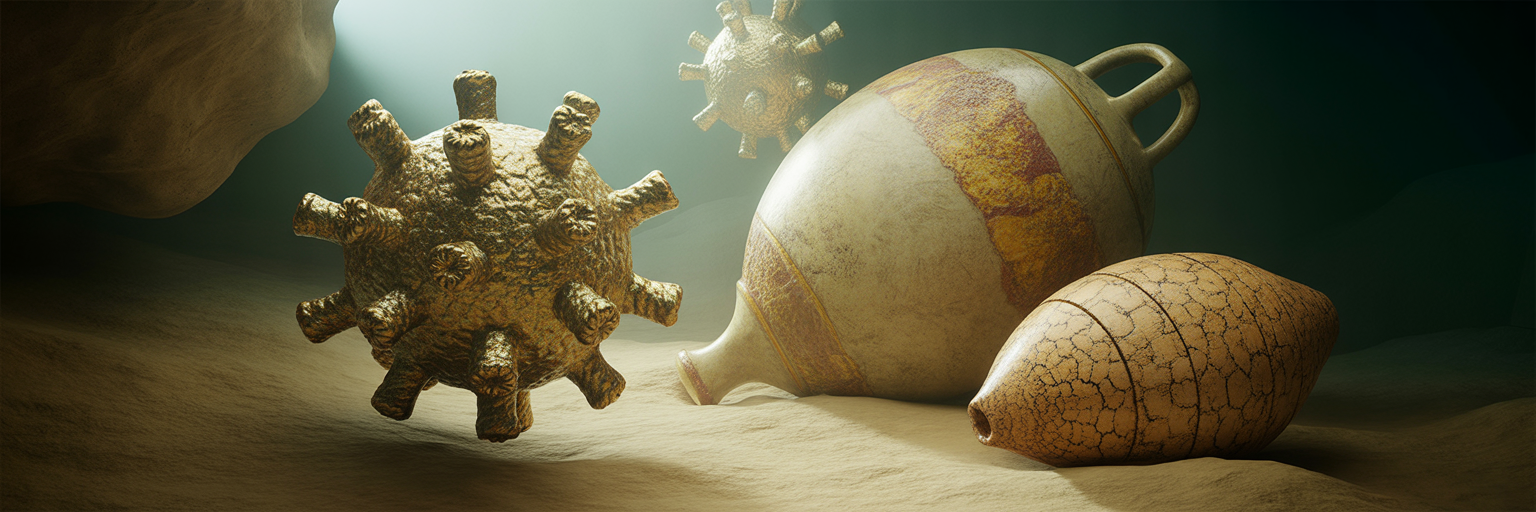
Soon after Mimivirus, the “Megavirus” family was identified in coastal waters off Chile, boasting an even larger genome. Then came the discovery of Pandoravirus, found in marine sediment and a freshwater pond in Australia. The name Pandoravirus reflects the surprise its discovery generated, as its unique amphora-like shape and colossal genome opened a new box of questions. At up to 2.5 million base pairs, its genome is larger than that of some parasitic eukaryotes, and over 90% of its genes are ORFans, unlike anything else in the known biological world.
Perhaps the most headline-grabbing discovery was Pithovirus sibericum, a permafrost virus isolated from a 30,000-year-old sample of Siberian ice. At 1.5 micrometers in length, it is the largest giant virus found to date. Its discovery proved that these viruses could remain viable for geological timescales in extreme environments. Other notable family members include the Tupanvirus, found in Brazil, which possesses the most complete set of protein-synthesis genes ever seen in a virus, and Mollivirus sibericum, another ancient virus recovered from the same Siberian permafrost sample.
This growing family shows that giant viruses are not anomalies but a widespread and diverse group of microbes found in oceans, freshwater sediments, soil, and even ancient ice. Their ability to persist in such varied and extreme conditions is a testament to their resilience. In the broader natural world, some animals have developed equally extraordinary methods for enduring harsh conditions, like the animal that survives by shrinking its own organs to conserve energy. The survival strategies of giant viruses are just as remarkable, pushing the boundaries of what we thought possible for a microbe.
Are Giant Viruses Alive? The Unresolved Debate
The existence of giant viruses has reignited one of biology’s most profound and contentious questions: what is the definition of life? Their size, genetic complexity, and semi-autonomous replication factories challenge the traditional boundary between chemistry and biology. As detailed in a 2024 review in Frontiers in Microbiology, the discovery of giant viruses has led to “the fall of an old paradigm,” forcing a continuous re-evaluation of these fundamental biological questions. Currently, there is no scientific consensus, with credible researchers arguing passionately on both sides.
The Case for ‘Non-Living’
The traditionalist view holds that viruses, including giant ones, are not alive. This argument focuses on the virion, the virus particle itself as it exists outside a host. In this state, it is metabolically inert. It cannot produce energy, it cannot synthesize its own proteins because it lacks ribosomes, and it cannot replicate without the machinery of a host cell. From this perspective, a virus is an intricate biochemical machine, but it lacks the autonomy that defines life. It is entirely dependent on a living cell to execute its genetic program.
The Case for ‘Living’
Proponents of the “living” virus hypothesis, including pioneers like Claverie and Abergel, argue that one should consider the entire viral life cycle, not just the inert particle. They point to the virus factory that forms inside the host cell. This structure actively recruits materials from the host, synthesizes viral components, and assembles new virions in a directed, organized fashion. They argue that this factory behaves like a temporary, living organism, a cell-like entity that uses the host as a source of raw materials. In this view, the virion is merely the dispersal stage, analogous to a seed or spore.
The Fourth Domain Hypothesis
The debate over whether giant viruses are alive is tied to the mystery of their origin. One leading theory, the reductive evolution hypothesis, proposes that giant viruses are descendants of a complex, ancient cellular ancestor. This ancestor may have belonged to a now-extinct fourth domain of life, alongside Bacteria, Archaea, and Eukarya. Over eons of parasitic existence, it gradually shed genes for independent life, such as those for ribosomes and energy production, while retaining a large and complex core genome. If true, giant viruses would be highly degenerated life forms, not complex non-life.
The Gene Accumulation Hypothesis
The competing theory suggests the opposite trajectory. In this scenario, giant viruses started as small, simple viruses and grew larger and more complex over time. Through a process called horizontal gene transfer, they gradually acquired genes from their hosts and other microbes in their environment. This would make them an extreme example of viral evolution, masters of genetic theft that assembled their complex genomes piece by piece. The profound questions they raise about life’s boundaries are similar to those posed by other strange biological phenomena, such as the secrets of planarian regeneration, where an organism can regrow its entire body from a fragment.
| Feature | Reductive Evolution Hypothesis | Gene Accumulation Hypothesis |
|---|---|---|
| Starting Point | A complex, ancient cellular ancestor | A small, simple ancestral virus |
| Primary Evolutionary Process | Gradual loss of genes for independent life | Gradual acquisition of genes from hosts |
| Source of Genes | Inherited from a cellular ancestor | Horizontal gene transfer from other organisms |
| Implication for Tree of Life | Represents a lost ‘fourth domain’ of life | Represents a highly evolved branch of viruses |
Human Health and Viruses from the Permafrost

The discovery of a 30,000-year-old permafrost virus that could still function created sensational headlines about “zombie viruses” thawing and threatening humanity. However, the scientific reality is far more nuanced. The study on Pithovirus sibericum demonstrated that an ancient virus could remain infectious, but it was only shown to be infectious to its specific host, an amoeba, under controlled laboratory conditions. It was not “revived” in the wild, and there is no evidence that it or other ancient giant viruses pose a direct threat to humans.
The key to understanding giant virus ecology is their relationship with amoebae. These single-celled protists are their primary natural hosts. Amoebae feed by engulfing large particles like bacteria in a process called phagocytosis. Because giant viruses are similar in size to bacteria, amoebae often “mistake” them for food and consume them. This provides the virus with a perfect entry point into its host. This specialized host-parasite relationship is a crucial part of their life cycle. Understanding why parasites need multiple hosts to party can offer insights into these complex ecological webs.
So, can giant viruses infect humans? The evidence is currently ambiguous. Traces of giant virus DNA have been detected in human samples, sometimes in patients with pneumonia, and have also been found on contact lenses. However, it is critical to distinguish between correlation and causation. The presence of viral DNA does not prove that the virus is causing disease. It could be a harmless passenger, an incidental finding, or even a contaminant from the environment. To date, no giant virus has been definitively proven to be a human pathogen. While the direct risk from the ancient permafrost virus is considered extremely low, the study serves as an important proof-of-concept. It shows that ancient microbes can retain viability over millennia, a finding of significant interest as climate change accelerates the thawing of permafrost.
A Quick Guide to Giant Viruses (FAQ)
The study of giant viruses has reshaped our understanding of the microbial world. Here are answers to some common questions about these fascinating entities.
How big is a giant virus?
Giant viruses are defined by their ability to be seen with a light microscope. Their physical size can range from about 400 nanometers to 1,500 nanometers (1.5 micrometers). Their genomes are also massive, ranging from hundreds of thousands to over 2.5 million base pairs, making them genetically more complex than many bacteria.
Can you see a giant virus with a normal microscope?
Yes. Unlike conventional viruses, which require a powerful electron microscope to be seen, giant viruses are large enough to be visible under a standard light microscope. This is why the first one, Mimivirus, was mistaken for a bacterium for over a decade.
Are giant viruses alive?
This is one of the most significant unresolved debates in biology. There is no consensus. Some scientists argue they are non-living because their particles are inert and they depend on a host for replication. Others argue they are living, considering their active and complex replication cycle inside a host cell. The question of are giant viruses alive remains open.
Can giant viruses infect humans?
Currently, there is no conclusive proof that giant viruses cause disease in humans. Their DNA has been found in human tissue samples, but a direct causal link has not been established. Their natural hosts are primarily single-celled organisms like amoebae.
Where are giant viruses found?
Giant viruses are ubiquitous and have been discovered in a wide range of environments across the globe. They have been found in ocean water, freshwater sediments, soil, desert environments, and even in 30,000-year-old Siberian permafrost.
Why do giant viruses have so many genes?
There are two main competing theories. The first is that they evolved from a more complex cellular ancestor and have lost genes over time (reductive evolution). The second is that they started as simple viruses and gradually accumulated genes from their hosts and other organisms (gene accumulation). This question is central to understanding their mysterious origins.